That's really great idea - to make 1 minute video story about stem cells. I like it a lot:
Showing posts with label basics. Show all posts
Showing posts with label basics. Show all posts
9/01/2015
7/24/2015
Let's create useful application for cell culturing!
In previous post I've asked about the necessity of mobile application development.
Now let's talk more specific.
While working with cell cultures I always notice lots of possibilities to improve the performance and make all procedures easier and to avoid mistakes.
However sometimes it's really hard to manage all cell cultures and to have all data available anywhere anytime.
That's why once I found myself thinking about the creation of something handy, trendy and highly useful for people who need to spend lots of time in laboratory.
My current idea is to develop the application for smartphones that will simplify some routine steps in cell cultivation process.
I'm not an application developer and I don't have enough funding to hire a good one today. That is why I've launched the campaign for this goal. Moreover, I'd like to hear any comments, wishes and recommendations from both cell scientists and people who know how such stuff as mobile app development works. It's important because I'd like to give the most precise description of this task to engineers.
Here's the donation page
All features are still under construction but here are some crucial goals that I want to see in ready-to-use app:
- Interactive Journal with the opportunity to put data abot the culture: isolation day (for primary cultures), seeding day (for other), number and date of passage, medium used (chose from given options e.g.: MEM, DMEM, RPMI etc), serum used (type of serum and %, e.g.: FBS, 10%), medium additives presence such as glutamine, glucose etc, the initial number of cells, type of flask used and other nesessary characteristics.
- This Journal will provide notifications - reminders of a regular medium change, subculturing etc
- Tools for cell counting such as population doubling time calculator, plotting options etc.
- One of my wishes is to make some sort of color detector that will give the possibility to determine the pH of the medium based on color on the photo
- Maybe there would be tools for image processing with microscope (linked to existing programs or connected with new simplified mobile programm)
- The possibility of additional (or only) marking of the culture flask using a code with information that appears in a smartphone
- Maybe a tool for rapid qualitative detection of signs of contamination by turbidity, color, environment
- An analytical package for the interpretation of test results (PRO)
Of course it's just a raw draft and you may also take part in creative process with your vision.
Thanks for all comments. It's really important!
7/23/2015
Apps for stem cell culturing
Dear readers, do you know any relevant stem cell related applications for smartphones?
Or maybe applications for assisting in cell culturing process?
And what do you think about the utility of such applications?
Thank you!
Or maybe applications for assisting in cell culturing process?
And what do you think about the utility of such applications?
Thank you!
1/13/2015
Wonderful stem cell blog
Just discovered the wonderful blog about the hot cell therapy topic! http://roosterbio.blogspot.com - there are lots of opinions on mesenchymal stem cell biology, clinical utility and other aspects. The most stunning thing about this blog is the courage for discussing the "uncomfortable" questions. These questions are intensively avoided because of some "anti-commercial" conclusions, but the resolving of such problems is extremely important for the development of safe and effective regenerative medicine.
12/24/2014
Umbilical Cord Tissue-Derived Cells as Therapeutic Agents
Olga Maslova, Miroslav Novak, and Peter Kruzliak, “Umbilical Cord Tissue-Derived Cells as Therapeutic Agents,” Stem Cells International, Article ID 150609, in press.
http://www.hindawi.com/journals/sci/aa/150609/ - our new work in Stem Cells International. I'm in love with this type of stem cells but need to admit that there are still lots of problems. I hope this paper would be useful for you.
http://www.hindawi.com/journals/sci/aa/150609/ - our new work in Stem Cells International. I'm in love with this type of stem cells but need to admit that there are still lots of problems. I hope this paper would be useful for you.
Abstract
Although the characteristics of SC, including UC-derived cells, are a dramatically discussed issue, this review will focus particularly on some controversial issues regarding clinical utility of cells isolated from UC tissue. UC-derived cells have several advantages compared to other types and sources of stem cells. The impact of UC topography on cell characteristics is briefly discussed. The necessity to adapt existing methods of cell isolation and culturing to GMP conditions is mentioned, as well as possible cryopreservation of this material. Light is shed on some future perspectives for UC-derived cells.
8/08/2014
2/15/2013
12/20/2012
positive interim safety results of its Phase IIa study of its allogeneic stem cell therapy
Cell Therapy pioneer TiGenix has announced positive interim safety results of its Phase IIa study of its allogeneic stem cell therapy Cx611 in rheumatoid arthritis (RA), showing a good safety profile at all three doses of the product that were administered in the trial.http://www.sciencebusiness.net/news/75982/European-cell-therapy-pioneer-makes-further-advance?utm_source
This is a significant development in the field of stem cell therapy because Cx611 is an allogeneic product that potentially could be made available off-the-shelf and used to treat any patient, without concerns that the foreign cells will cause an immune reaction. In addition, TiGenix says the product, which is made from adult stem cells derived from human adipose (fat) tissue, has a broad anti-inflammatory effect and could also be used to treat other autoimmune disorders.
If positive, the final results of the trial, due in April 2013, will set the scene for the further development of Cx611. The TiGenix trial is the most advanced in the world using stem cells to treat rheumatoid arthritis. Along with other cell therapies that are advancing in development such as ReNeuron plc’s neuronal cell therapy for treating the after-effects of stroke, and the human embryonic stem cell-based treatment for macular degeneration (a major cause of blindness) which Pfizer is developing in collaboration with scientists at University College London, the TiGenix news on Cx611 underlines Europe’s leading position in cell therapy and regenerative medicine.
These three products can all trace their origins back to publicly-funded basic research, providing a potent demonstration of the importance of continuing to support academic research in the field, and of building and reinforcing the clinical and regulatory framework for translating this research into commercial products.
Moves by some MEPs to end European Union funding for embryonic stem cell research in the proposed €80 billion Horizon 2020 R&D programme are widely seen as a threat to Europe’s standing in cell therapy and regenerative medicine. Science|Business brought together experts from patients’ groups, research charities, academe, industry, science and economic policy, and regulators, to discuss the implications of an end to EU support for embryonic stem cell research, and scope what needs to be done to build the regenerative medicines market in Europe, to the benefit of patients and the economy. The full report is now available here.
8/27/2012
.
Here is the express Stem Cell news on the other place - my paper.li. Very interesting place to put your information. I like it!:
5/26/2012
Stem Cell Comics
Open publication - Free publishing
Beautiful educational comics about stem cells! ‘Hope Beyond Hype’ is a story about stem cell therapies from science discovery to working therapy.
4/19/2012
Beautiful Cell Pictures
Those beautiful cell pictures were found at
http://bpod.mrc.ac.uk/search?utf8=%E2%9C%93&keywords=stem+cells


2/24/2012
Stem Cell Aging
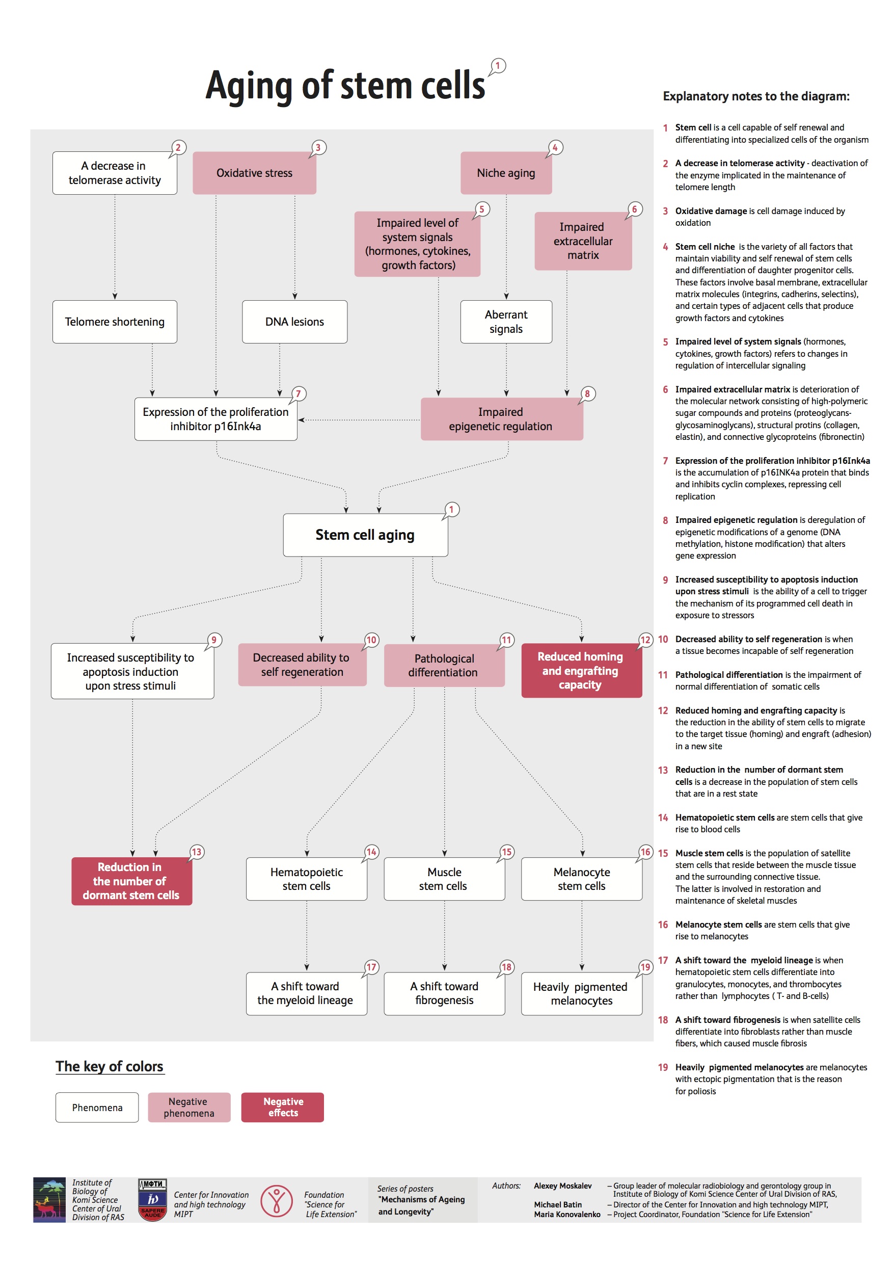
http://www.fightaging.org/images/aging_of_stem_cells.jpg
the problem of Stem Cell Aging is one of the most intrigueing in stem cell biology field.
1/20/2012
Stem Cell Research Problems
Another one opinion on stem cell research:
http://shyfish.info/a-peek-at-each-side-in-the-stem-cell-studies-discussion/
A lot has been written and also said about stem cell research, and most of these trumpet about ultimately locating a cure for several ailments. Nevertheless, presently there are also camps that absolutely dismiss the stem cell research pros, while focusing on its purportedly damaging aspects. But before many of us live on the stem cell research disadvantages, allow us very first discuss exactly what come cellular material are regarding. Originate cells produce fresh blood vessels tissues, which is why they will are considered as the blood’s human being building blocks. The thing that makes come cells particular is the simple fact that they can possibly become various kinds of cells, which usually in switch may be developed into specialized cellular material that may be used by scientists in their own search for ways to repair bodily organs that have by now sustained harm.
Of the a couple of vast forms of stem tissues observed in mammals, embryonic base cells are the versions that could possibly grow into other kinds of tissues. Very easily stored in a laboratory, embryonic base cellular material, are additionally special in the feeling that they are able to identical them selves in countless amounts, along with forever with that.
This quality on your own means they are ideal for scientific utilize along with ongoing stem cell research. Most of this reports have already proven that serious diseases similar to ms, Parkinson's along with Alzheimer's is treatable with the assistance of come tissue. That perhaps retains the potential to the treatment of heart disease, strokes, kidney disease, liver disease, pancreas disease and also arthritis, as well as discovering a treat for cancer malignancy.
But for almost all the stem cell research pros, some groupings notice the concerns in the idea. Much of the stem cell research downsides that that they are professing are moral in character. These people say that because embryonic come cells are in truth little embryos, just what stem mobile researchers are tinkering with is living. Almost all of those who take this distinct argument are those which consider that existence starts with conception, certainly not at start, as just what is typically accepted.
The problem of cloning has also hounded stem cellular scientists, which profess they only rely on them for research functions, not necessarily to clone another individual. Nevertheless whichever the problems for or versus stem cell research are, the truth remains that teens is on the verge involving a main health-related as well as scientific jump, one that can modify the globe once and for all.
1/10/2012
1/09/2012
Education (stem cell topic)
Beautiful stem cell education resource: http://www.tellmeaboutstemcells.org/
The Best Mesenchymal Stem Cell Reviews (part 1)
I'm involved in MSC-research and I love brilliant articles on that topic. The list of my favorite reviews about Mesenchymal Stem Cells is here now:
 Klopp, A., Gupta, A., Spaeth, E., Andreeff, M., & Marini, F. (2011). Concise Review: Dissecting a Discrepancy in the Literature: Do Mesenchymal Stem Cells Support or Suppress Tumor Growth? STEM CELLS, 29 (1), 11-19 DOI: 10.1002/stem.559
KNEE SURGERY, SPORTS TRAUMATOLOGY, ARTHROSCOPY
Volume 17, Number 11, 1289-1297, DOI: 10.1007/s00167-009-0782-4
A Comprehensive Review on Mesenchymal Stem Cell Growth and Senescence Krzysztof Książek. Rejuvenation Research. April 2009, 12(2): 105-116. doi:10.1089/rej.2009.0830.
Kim, S., & de Vellis, J. (2009). Stem cell-based cell therapy in neurological diseases: A review Journal of Neuroscience Research, 87 (10), 2183-2200 DOI: 10.1002/jnr.22054
Stem Cells Trans MedDecember 2011sctm.2011-0024
Concise Review: Clinical Translation of Wound Healing Therapies Based on Mesenchymal Stem Cells
Wesley M. Jackson, Leon J. Nesti, and Rocky S. Tuan
Inflammation and mesenchymal stem cell aging Gunter Lepperdinger
Current Opinion in Immunology Volume 23, Issue 4, August 2011, Pages 518-524
doi:10.1016/j.coi.2011.05.007
Karp, J., & Leng Teo, G. (2009). Mesenchymal Stem Cell Homing: The Devil Is in the Details Cell Stem Cell, 4 (3), 206-216 DOI: 10.1016/j.stem.2009.02.001
To be continued...
KNEE SURGERY, SPORTS TRAUMATOLOGY, ARTHROSCOPY
Volume 17, Number 11, 1289-1297, DOI: 10.1007/s00167-009-0782-4
Stem Cells Trans MedDecember 2011sctm.2011-0024
Concise Review: Clinical Translation of Wound Healing Therapies Based on Mesenchymal Stem Cells
Wesley M. Jackson, Leon J. Nesti, and Rocky S. Tuan
Inflammation and mesenchymal stem cell aging Gunter Lepperdinger
Current Opinion in Immunology Volume 23, Issue 4, August 2011, Pages 518-524
doi:10.1016/j.coi.2011.05.007
And the very best:
To be continued...
1/06/2012
Stemmy!111 ^_^
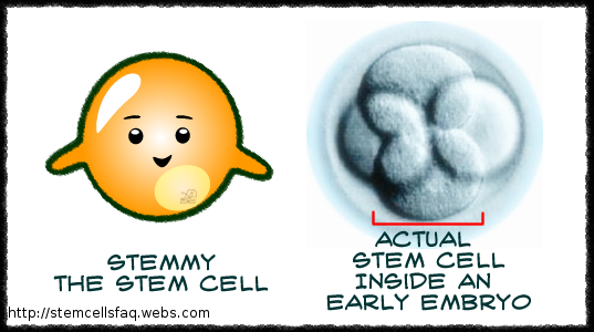
And more about Stemmy:
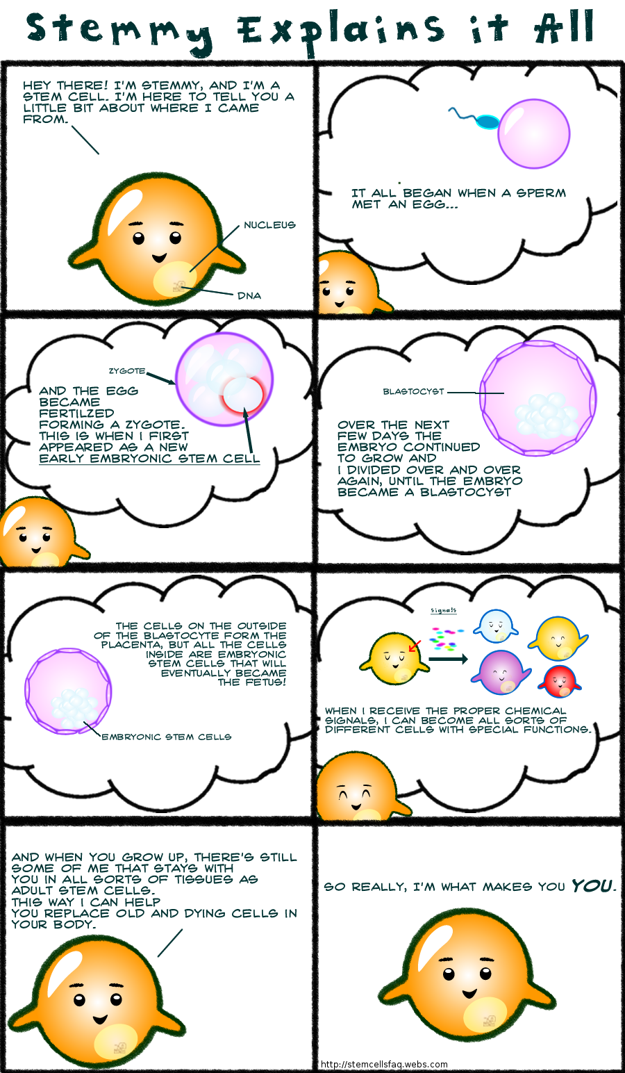
Aaaaaannnnnddddd moooooooreeee:
Pictures from http://www.terry.ubc.ca/2009/08/07/faq-stem-cells-sa-mix/, moreover you can find text there.
12/29/2011
12/27/2011
Stem Cells!
Beautiful Stem Cells! Thnx to
http://stemcelldaily.com/stem-cells-are-purdy-pics/ :

Neurosphere composed of neural precursor cells as captured by a fluorescent microscope. The cells, allowed to attach to a substrate, have begun to send out long processes that will eventually become the axons of the mature neurons.
The image was taken in the lab of Martin Pera at the University of Southern California.

A confocal microscopic image of a neurosphere, a ball of human embryonic stem cells giving rise to nerve cells. The nuclei of the neurons are shown in blue, while the axons are shown in red.
The image was taken in the lab of Juan Carlos Izpisua Belmonte at The Salk Institute for Biological Studies.

Color-enhanced electron microscopic image of mouse embryonic stem cells growing on a bed of silicon nanotubes.
The image was taken in the lab of Bruce Conklin at the Gladstone Institute for Cardiovascular Medicine.

Color-enhanced image taken by a scanning electron microscope of retinal pigment epithelial (RPE) cells derived from human embryonic stem cells. The cells are remarkably similar to normal RPE cells, having a hexagonal shape and growing in a single, well defined layer. These cells are the ones responsible for macular degeneration, the most common cause of blindness. CIRM scientists hope to one day treat macular degeneration with transplanted RPE cells derived from human embryonic stem cells.
The image was taken in the lab of David Hinton at the University of Southern California.
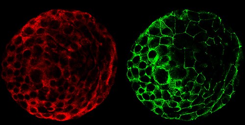

A composite of two images taken of a human embryo under different fluorescent wavelengths using a confocal microscope.
Fluorescent tags reveal that cells on the surface of the embryo are expressing human chorinoic gonadotropin (green tag) and an adhesion molecule (red tag)that helps them stick together.
The image was taken in the lab of Susan Fisher at the University of California, San Francisco.
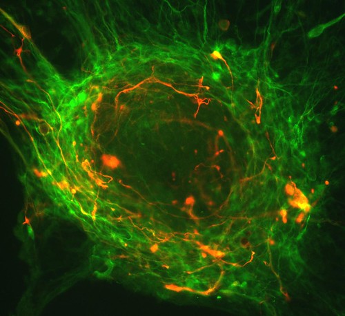
A fluorescent microscopic image of hundreds of human embryonic stem cells in various stages of differentiation into neurons. Some cells have become neurons (red), while others are still precursors of nerve cells (green). The yellow is an imaging artifact that results when cells in both stages are on top of each other.
The image was taken in the lab of Guoping Fan at the University of California, Los Angeles.

Colonies of human embryonic stem cells as seen with a fluorescent microscope. Nuclei have been stained blue, while regions that appear pink and green have been stained with antibodies, indicating the cells’ pluripotency — that unique ability of stem cells to differentiate into a variety of cell types.
The image was taken in the lab of Prue Talbot at the University of California, Riverside.

Three neurons (red) derived from human embryonic stem cells (hESCs) as seen by a confocal microscope. Visible are neural cell bodies, complete with axons and dendrites (red), used for cell-to-cell communications, as well as undifferentiated hESCs (green).
The image was taken in the lab of Anirvan Ghosh at the University of California, San Diego.

A fluorescent microscopic image of a functional neuron with an axon (red) growing above the cell’s nucleus and three dendrites (red) below. Undifferentiated neural precursor cells (blue) are visible as are glia cells (green) that have differentiated from the same group of mouse neural stem cells.
The image was taken in the lab of Paul Knoepfler at the University of California, Davis.

Two neurospheres, compact masses of neuron precursor cells, derived from human embryonic stem cells, as captured by a fluorescent microscope. Differentiated neurons, whose nuclei are shown in red, have begun to extend neuronal processes (green) toward one another, forming connections.
Neurosphere composed of neural precursor cells as captured by a fluorescent microscope. The cells, allowed to attach to a substrate, have begun to send out long processes that will eventually become the axons of the mature neurons.
The image was taken in the lab of Martin Pera at the University of Southern California.
A confocal microscopic image of a neurosphere, a ball of human embryonic stem cells giving rise to nerve cells. The nuclei of the neurons are shown in blue, while the axons are shown in red.
The image was taken in the lab of Juan Carlos Izpisua Belmonte at The Salk Institute for Biological Studies.
Color-enhanced electron microscopic image of mouse embryonic stem cells growing on a bed of silicon nanotubes.
The image was taken in the lab of Bruce Conklin at the Gladstone Institute for Cardiovascular Medicine.
Color-enhanced image taken by a scanning electron microscope of retinal pigment epithelial (RPE) cells derived from human embryonic stem cells. The cells are remarkably similar to normal RPE cells, having a hexagonal shape and growing in a single, well defined layer. These cells are the ones responsible for macular degeneration, the most common cause of blindness. CIRM scientists hope to one day treat macular degeneration with transplanted RPE cells derived from human embryonic stem cells.
The image was taken in the lab of David Hinton at the University of Southern California.
A composite of two images taken of a human embryo under different fluorescent wavelengths using a confocal microscope.
Fluorescent tags reveal that cells on the surface of the embryo are expressing human chorinoic gonadotropin (green tag) and an adhesion molecule (red tag)that helps them stick together.
The image was taken in the lab of Susan Fisher at the University of California, San Francisco.
A fluorescent microscopic image of hundreds of human embryonic stem cells in various stages of differentiation into neurons. Some cells have become neurons (red), while others are still precursors of nerve cells (green). The yellow is an imaging artifact that results when cells in both stages are on top of each other.
The image was taken in the lab of Guoping Fan at the University of California, Los Angeles.
Colonies of human embryonic stem cells as seen with a fluorescent microscope. Nuclei have been stained blue, while regions that appear pink and green have been stained with antibodies, indicating the cells’ pluripotency — that unique ability of stem cells to differentiate into a variety of cell types.
The image was taken in the lab of Prue Talbot at the University of California, Riverside.
Three neurons (red) derived from human embryonic stem cells (hESCs) as seen by a confocal microscope. Visible are neural cell bodies, complete with axons and dendrites (red), used for cell-to-cell communications, as well as undifferentiated hESCs (green).
The image was taken in the lab of Anirvan Ghosh at the University of California, San Diego.
A fluorescent microscopic image of a functional neuron with an axon (red) growing above the cell’s nucleus and three dendrites (red) below. Undifferentiated neural precursor cells (blue) are visible as are glia cells (green) that have differentiated from the same group of mouse neural stem cells.
The image was taken in the lab of Paul Knoepfler at the University of California, Davis.
Two neurospheres, compact masses of neuron precursor cells, derived from human embryonic stem cells, as captured by a fluorescent microscope. Differentiated neurons, whose nuclei are shown in red, have begun to extend neuronal processes (green) toward one another, forming connections.
The image was taken in the lab of Fred H. Gage at the Salk Institute for Biological Studies.

Neurospheres made up of neural stem cells derived from human embryonic stem cells captured using fluorescence microscopy. Some cells (green) are destined to become neurons; others have yet to differentiate (red) or are in transition (yellow).
This photo was taken in the lab of Brian Cummings at the University of California, Irvine.

A composite image of frames taken of a developing human embryo captured using time-lapse video microscopy and a phase contrast microscope. The earliest frame is of a three-day-old embryo and appears in the upper left corner. An image of a five-day-old embryo appears in the lower right.
This photo was taken in the lab of Susan Fisher at the University of California, San Francisco.
You can find out more information about the contest here.
Neurospheres made up of neural stem cells derived from human embryonic stem cells captured using fluorescence microscopy. Some cells (green) are destined to become neurons; others have yet to differentiate (red) or are in transition (yellow).
This photo was taken in the lab of Brian Cummings at the University of California, Irvine.
A composite image of frames taken of a developing human embryo captured using time-lapse video microscopy and a phase contrast microscope. The earliest frame is of a three-day-old embryo and appears in the upper left corner. An image of a five-day-old embryo appears in the lower right.
This photo was taken in the lab of Susan Fisher at the University of California, San Francisco.
You can find out more information about the contest here.
Subscribe to:
Posts (Atom)

