Beautiful Stem Cells! Thnx to
http://stemcelldaily.com/stem-cells-are-purdy-pics/ :

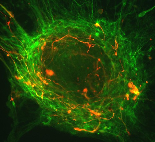
Neurosphere composed of neural precursor cells as captured by a fluorescent microscope. The cells, allowed to attach to a substrate, have begun to send out long processes that will eventually become the axons of the mature neurons.
The image was taken in the lab of Martin Pera at the University of Southern California.

A confocal microscopic image of a neurosphere, a ball of human embryonic stem cells giving rise to nerve cells. The nuclei of the neurons are shown in blue, while the axons are shown in red.
The image was taken in the lab of Juan Carlos Izpisua Belmonte at The Salk Institute for Biological Studies.

Color-enhanced electron microscopic image of mouse embryonic stem cells growing on a bed of silicon nanotubes.
The image was taken in the lab of Bruce Conklin at the Gladstone Institute for Cardiovascular Medicine.

Color-enhanced image taken by a scanning electron microscope of retinal pigment epithelial (RPE) cells derived from human embryonic stem cells. The cells are remarkably similar to normal RPE cells, having a hexagonal shape and growing in a single, well defined layer. These cells are the ones responsible for macular degeneration, the most common cause of blindness. CIRM scientists hope to one day treat macular degeneration with transplanted RPE cells derived from human embryonic stem cells.
The image was taken in the lab of David Hinton at the University of Southern California.
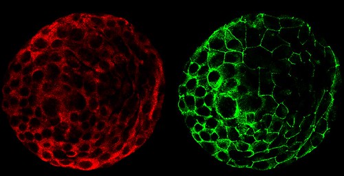

A composite of two images taken of a human embryo under different fluorescent wavelengths using a confocal microscope.
Fluorescent tags reveal that cells on the surface of the embryo are expressing human chorinoic gonadotropin (green tag) and an adhesion molecule (red tag)that helps them stick together.
The image was taken in the lab of Susan Fisher at the University of California, San Francisco.
A fluorescent microscopic image of hundreds of human embryonic stem cells in various stages of differentiation into neurons. Some cells have become neurons (red), while others are still precursors of nerve cells (green). The yellow is an imaging artifact that results when cells in both stages are on top of each other.
The image was taken in the lab of Guoping Fan at the University of California, Los Angeles.

Colonies of human embryonic stem cells as seen with a fluorescent microscope. Nuclei have been stained blue, while regions that appear pink and green have been stained with antibodies, indicating the cells’ pluripotency — that unique ability of stem cells to differentiate into a variety of cell types.
The image was taken in the lab of Prue Talbot at the University of California, Riverside.

Three neurons (red) derived from human embryonic stem cells (hESCs) as seen by a confocal microscope. Visible are neural cell bodies, complete with axons and dendrites (red), used for cell-to-cell communications, as well as undifferentiated hESCs (green).
The image was taken in the lab of Anirvan Ghosh at the University of California, San Diego.

A fluorescent microscopic image of a functional neuron with an axon (red) growing above the cell’s nucleus and three dendrites (red) below. Undifferentiated neural precursor cells (blue) are visible as are glia cells (green) that have differentiated from the same group of mouse neural stem cells.
The image was taken in the lab of Paul Knoepfler at the University of California, Davis.

Two neurospheres, compact masses of neuron precursor cells, derived from human embryonic stem cells, as captured by a fluorescent microscope. Differentiated neurons, whose nuclei are shown in red, have begun to extend neuronal processes (green) toward one another, forming connections.
Neurosphere composed of neural precursor cells as captured by a fluorescent microscope. The cells, allowed to attach to a substrate, have begun to send out long processes that will eventually become the axons of the mature neurons.
The image was taken in the lab of Martin Pera at the University of Southern California.
A confocal microscopic image of a neurosphere, a ball of human embryonic stem cells giving rise to nerve cells. The nuclei of the neurons are shown in blue, while the axons are shown in red.
The image was taken in the lab of Juan Carlos Izpisua Belmonte at The Salk Institute for Biological Studies.
Color-enhanced electron microscopic image of mouse embryonic stem cells growing on a bed of silicon nanotubes.
The image was taken in the lab of Bruce Conklin at the Gladstone Institute for Cardiovascular Medicine.
Color-enhanced image taken by a scanning electron microscope of retinal pigment epithelial (RPE) cells derived from human embryonic stem cells. The cells are remarkably similar to normal RPE cells, having a hexagonal shape and growing in a single, well defined layer. These cells are the ones responsible for macular degeneration, the most common cause of blindness. CIRM scientists hope to one day treat macular degeneration with transplanted RPE cells derived from human embryonic stem cells.
The image was taken in the lab of David Hinton at the University of Southern California.
A composite of two images taken of a human embryo under different fluorescent wavelengths using a confocal microscope.
Fluorescent tags reveal that cells on the surface of the embryo are expressing human chorinoic gonadotropin (green tag) and an adhesion molecule (red tag)that helps them stick together.
The image was taken in the lab of Susan Fisher at the University of California, San Francisco.
A fluorescent microscopic image of hundreds of human embryonic stem cells in various stages of differentiation into neurons. Some cells have become neurons (red), while others are still precursors of nerve cells (green). The yellow is an imaging artifact that results when cells in both stages are on top of each other.
The image was taken in the lab of Guoping Fan at the University of California, Los Angeles.
Colonies of human embryonic stem cells as seen with a fluorescent microscope. Nuclei have been stained blue, while regions that appear pink and green have been stained with antibodies, indicating the cells’ pluripotency — that unique ability of stem cells to differentiate into a variety of cell types.
The image was taken in the lab of Prue Talbot at the University of California, Riverside.
Three neurons (red) derived from human embryonic stem cells (hESCs) as seen by a confocal microscope. Visible are neural cell bodies, complete with axons and dendrites (red), used for cell-to-cell communications, as well as undifferentiated hESCs (green).
The image was taken in the lab of Anirvan Ghosh at the University of California, San Diego.
A fluorescent microscopic image of a functional neuron with an axon (red) growing above the cell’s nucleus and three dendrites (red) below. Undifferentiated neural precursor cells (blue) are visible as are glia cells (green) that have differentiated from the same group of mouse neural stem cells.
The image was taken in the lab of Paul Knoepfler at the University of California, Davis.
Two neurospheres, compact masses of neuron precursor cells, derived from human embryonic stem cells, as captured by a fluorescent microscope. Differentiated neurons, whose nuclei are shown in red, have begun to extend neuronal processes (green) toward one another, forming connections.
The image was taken in the lab of Fred H. Gage at the Salk Institute for Biological Studies.

Neurospheres made up of neural stem cells derived from human embryonic stem cells captured using fluorescence microscopy. Some cells (green) are destined to become neurons; others have yet to differentiate (red) or are in transition (yellow).
This photo was taken in the lab of Brian Cummings at the University of California, Irvine.

A composite image of frames taken of a developing human embryo captured using time-lapse video microscopy and a phase contrast microscope. The earliest frame is of a three-day-old embryo and appears in the upper left corner. An image of a five-day-old embryo appears in the lower right.
This photo was taken in the lab of Susan Fisher at the University of California, San Francisco.
You can find out more information about the contest here.
Neurospheres made up of neural stem cells derived from human embryonic stem cells captured using fluorescence microscopy. Some cells (green) are destined to become neurons; others have yet to differentiate (red) or are in transition (yellow).
This photo was taken in the lab of Brian Cummings at the University of California, Irvine.
A composite image of frames taken of a developing human embryo captured using time-lapse video microscopy and a phase contrast microscope. The earliest frame is of a three-day-old embryo and appears in the upper left corner. An image of a five-day-old embryo appears in the lower right.
This photo was taken in the lab of Susan Fisher at the University of California, San Francisco.
You can find out more information about the contest here.


No comments:
Post a Comment