Intro

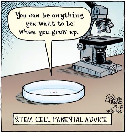
I work with stem cells in the lab, so I want to share the information on that topic at that blog. There are lots of interesting articles and study materials about stem cells on the web that's why I'm trying to gather them all here.

12/24/2014

Umbilical Cord Tissue-Derived Cells as Therapeutic Agents

Olga Maslova, Miroslav Novak, and Peter Kruzliak, “Umbilical Cord Tissue-Derived Cells as Therapeutic Agents,” Stem Cells International, Article ID 150609, in press.
http://www.hindawi.com/journals/sci/aa/150609/ - our new work in Stem Cells International. I'm in love with this type of stem cells but need to admit that there are still lots of problems. I hope this paper would be useful for you.
Abstract
Although the characteristics of SC, including UC-derived cells, are a dramatically discussed issue, this review will focus particularly on some controversial issues regarding clinical utility of cells isolated from UC tissue. UC-derived cells have several advantages compared to other types and sources of stem cells. The impact of UC topography on cell characteristics is briefly discussed. The necessity to adapt existing methods of cell isolation and culturing to GMP conditions is mentioned, as well as possible cryopreservation of this material. Light is shed on some future perspectives for UC-derived cells.

9/29/2014

Tonsil-Derived MSC

It was quite predictable and here it is:
"Polypeptide Thermogels as a Three Dimensional Culture Scaffold for Hepatogenic Differentiation of Human Tonsil-Derived Mesenchymal Stem Cells"
ACS Applied Materials & Interfaces
The liver provides critical functions, such as ridding the body of toxins. Its failure can be deadly, and there are few options for fixing it. But scientists now report in the journalACS Applied Materials & Interfaces a way to potentially inject stem cells from tonsils, a body part we don’t need, to repair damaged livers — all without surgery.
Byeongmoon Jeong and colleagues point out that currently, the only established method for treating liver failure or severe cases of liver disease is complete or partial transplantation. But the need is much greater than the number of available organs. Plus, surgery has inherent risks and a hefty price tag. A promising alternative in development is transplanting liver cells. One such approach involves using adult stem cells to make liver cells. Stem cells from bone marrow could be used, but they have limitations. Recently, scientists identified another source of adult stem cells that could be used for this purpose — tonsils. Every year, thousands of surgeries are performed to remove tonsils, and the tissue is discarded. Now it could have a new purpose, but scientists needed a way to grow them on a 3-D scaffold that mimics real liver tissue. Jeong’s team set out to do just that.
The researchers encapsulated tonsil-derived stem cells in a heat-sensitive liquid that turns into a gel at body temperature. They added substances called growth factors to encourage the stem cells to become liver cells. Then, they heated the combination up to a normal body temperature. The result was a 3-D, biodegradable gel that contained functioning liver cells. The researchers conclude that the same process has promise — with some further tweaking for ideal conditions — as an injectable tissue engineering technique to treat liver disease without surgery.
The authors acknowledge funding from the National Research Foundation of Korea.
I think it could be really possible to obtain stem cells from different tissues.
Thanks to http://www.acs.org/ for the information.

8/29/2014

Gremlins Sabotage the Mechanisms of Cancer Stem Cell Differentiation

Gremlins Sabotage the Mechanisms of Cancer Stem Cell Differentiation - that's not a joke. It's a title of the new paper at Cancer Cell Volume 25, Issue 6, 16 June 2014, Pages 716–717. Joan Seoane made an interesting research on cancer stem cells. 
Here is the abstract: BMP is highly expressed in glioblastoma and promotes differentiation of cancer stem cells (CSCs). Recently, Yan and colleagues found the explanation to this apparent paradox by showing that the antagonist of BMP, Gremlin1, is secreted by CSCs to protect them against the BMP-induced differentiation.

5/13/2013

Biomaterials for Stem Cell Therapy - CRC Press Book

Biomaterials for Stem Cell Therapy - CRC Press Book

Focused on stem cell applications, this book bridges the fields of biomaterials, offering new insights into constructing and regenerating tissues and organs. Its unique feature is linking diseases of the human body to current thinking on how to deal with them in the context of current concepts and technologies by means of an in-depth focus on biomaterials. The book assembles recent advances and covers a range of topics related to stem cell biology, biomaterials and technological approaches such as bioreactors written by top researchers in the field. Stem cells of both embryonic and adult origin are discussed with applications ranging, but not limited to, nerve regeneration, liver, pancreas, skin, trachea, cartilage and bone repair and cardiovascular therapy. Developments in the field reflecting the design and construction of the human body and its principal anatomy are discussed from a materials point of view.
The book will be a valuable tool for biomaterial scientists, tissue engineers, clinicians as well as stem cell biologists involved in basic research and applications of adult and embryonic stem cells. It will also be a source of reference for students in biotechnology, biomedical engineering, biology, biochemistry, materials sciences, pharmaceuticals, and veterinary and human medicine.

2/07/2013

3-D printers can produce gun parts, aircraft wings, food and a lot more, but this new 3-D printed product may be the craziest thing yet: human embryonic stem cells.

Rebecca Boyle - Popular Science

3-D printers can produce gun parts, aircraft wings, food and a lot more, but this new 3-D printed product may be the craziest thing yet: human embryonic stem cells. Using stem cells as the "ink" in a 3-D printer, researchers in Scotland hope to eventually build 3-D printed organs and tissues. A team at Heriot-Watt University used a specially designed valve-based technique to deposit whole, live cells onto a surface in a specific pattern.

This article originally appeared at Popular Science.

The cells were floating in a "bio-ink," to use the terminology of the researchers who developed this technique. They were able to squeeze out tiny droplets, containing five cells or fewer per droplet, in a variety of shapes and sizes. To produce clumps of cells, the team printed out cells first and then overlaid those with cell-free bio-ink, resulting in larger droplets or spheroids of cells. The cells would group together inside these spheroids. Spheroid size is key, because stem cells need certain conditions to work properly. This is why very precisely controlled 3-D printing could be so valuable for stem cell research.

After being squeezed out of a thin valve, the cells were still alive and viable, and able to transform into any other cell in the body, the researchers say. It's the first time anyone has printed human embyronic stem cells, said lead researcher Will Wenmiao Shu, a professor at Heriot-Watt. But ... why?

Eventually, they could be used to print out new tissues, or as filler inside existing organs, which would be regenerated. It could even serve to limit animal testing for new drug compounds, allowing them to be tested on actual human tissue, said Jason King, business development manager at Roslin Cellab, one of the research partners. "In the longer term, [it could] provide organs for transplant on demand, without the need for donation and without the problems of immune suppression and potential organ rejection," he said in a statement.

The team took stem cells from an embryonic kidney and from a well-studied embryonic cell line, and grew them in culture. They had to build a custom reservoir — let's call it an inkwell — to safely house the delicate cells, and then they added some large-diameter nozzles. A pressurized air supply pumps the cells from the inkwell into the valves, which contain pressurized nozzles on the end. The team could control the amount of cells dispensed by changing any of the factors, including the pneumatic pressure, nozzle diameter or length of time the nozzle stayed open.

At first the researchers printed droplets, but ultimately, they were so precise that they made cell spheroids in a variety of shapes and sizes, like the university logo above. One interesting wrinkle: The cells also formed spheroids in the inkwells. More work needs to be done to explain that.

The researchers also took several steps to make sure the cells survived the printing process. Examining the results of several experiments, they found 99 percent of the cells were still viable after running through the valve-based printer. "This confirms that this printing process did not appear to damage the cells or affect the viability of the vast majority of dispensed cells," they write in their paper, which is being published in the IOP regenerative medicine journal Biofabrication.

Stem cells are powerful because they can develop into any cell in the body. Embryonic stem cells, which are taken from human embryos in the earliest stages of development, can be developed into stem cell lines that can be grown indefinitely. This is kind of controversial, especially in the US. But medical researchers think they could be hugely promising for a whole host of human ailments — stem cells could differentiate into neurons, potentially replacing the ones lost in degenerative diseases like Alzheimer's; or they could differentiate into pancreatic cells, curing diabetes; and so on.

Using a 3-D printer to produce gun parts has been pretty controversial, especially during the ongoing post-Connecticut-shooting gun debate. But that may be nothing compared to this.

http://io9.com/5981832/a-3d-printer-that-generates-human-embryonic-stem-cells

1/26/2013

Leprosy & stem cells


Leprosy is a bacterial disease that spreads to muscles and other tissues in the body, causing neurodegeneration and muscle weakness. A new study, published by Cell Press January 17th in the journal Cell, reveals that the bacteria responsible for leprosy spread infection by hijacking specialized cells in the adult nervous system, reprogramming them into a stem cell-like state, and converting them to muscle-like cells. These findings could lead to the development of new therapeutic strategies for combating bacterial infections and degenerative diseases as well as new tools for regenerative medicine.
"This is the first demonstration of how a bacterial pathogen could use the genomic plasticity of our adult body tissue cells for generating stem cells naturally during infection," says senior study author Anura Rambukkana of the University of Edinburgh. "Our findings provide new directions for preventing the progression of infection at an early stage and for reprogramming adult tissue cells to stem cells for regenerating damaged tissues in the body."
Leprosy is caused by Mycobacterium leprae (M. leprae), which initially infects adult Schwann cells, cells which usually wrap around nerves to insulate electrical signals passing through, in the peripheral nervous system. The leprosy bacteria must then spread to other tissues to transmit infection, but how they do so has been a long-standing mystery. Because Schwann cells can convert into dedicated repair cells to help adult nerves recover after injury, Rambukkana and his team suspected that M. leprae takes advantage of this remarkable plasticity to spread infection.
To test this idea, the researchers infected adult Schwann cells from mice with M. leprae. The bacteria reprogrammed these cells into a stem cell-like state—in which they're capable of converting into diverse cell types—by turning off genes that are active in the mature form of these cells and turning on genes that are expressed during embryonic development. M. leprae then converted these immature cells into muscle-like cells and spread infection to muscles through this process. When the researchers injected bacteria-laden immature cells into the muscles of adult mice, the bacteria spread to different types of muscle cells.
"Our study shows that host cell reprogramming is perhaps a necessary event in early bacterial infection that promotes the spread of infection," Rambukkana says. "By identifying early molecular targets or diagnostic biomarkers related to the reprogramming process, it will be possible to prevent the progression of infection and thus nerve damage and subsequent disability in patients."

Cell, Masaki et al.: "Reprogramming Adult Schwann Cells to Stem Cell-Like Cells by Leprosy Bacilli Promotes Dissemination of Infection."
Interesting news! Thanks to  http://www.eurekalert.org/pub_releases/2013-01/cp-nii011013.php 
Elleven%20Project